Home
/ Picture Of The Human Spine - 17 593 Human Vertebra Stock Photos Pictures Royalty Free Images Istock, In some subjects, the seventh cervical vertebra is associated with an abnormal pair of ribs, known as cervical ribs.
Picture Of The Human Spine - 17 593 Human Vertebra Stock Photos Pictures Royalty Free Images Istock, In some subjects, the seventh cervical vertebra is associated with an abnormal pair of ribs, known as cervical ribs.
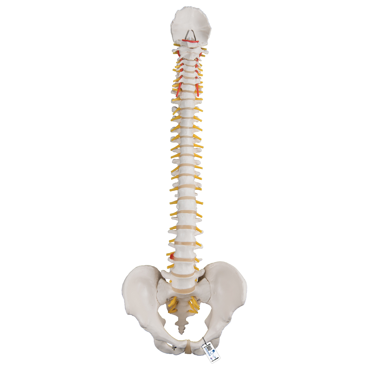
Picture Of The Human Spine - 17 593 Human Vertebra Stock Photos Pictures Royalty Free Images Istock, In some subjects, the seventh cervical vertebra is associated with an abnormal pair of ribs, known as cervical ribs.. These vertebrae bear much of the body's weight and related biomechanical stress. Five bones (abbreviated s1 through s5) fused into a triangular shape, form the sacrum. As part of the pelvic girdle, the sacrum forms the back wall of the pelvis and also forms joints at the hip bone called the sacroiliac joints. Little or no voluntary control of bowel or bladder, but patients can manage on their own with special equipment most likely they will also still be able to walk. The inner layer, or gray matter, is mainly composed of nerve cell bodies.
Injury to spinal bone three often causes pain, tingling, and sometimes numbness in the arms, neck, and head. Within the gray matter, running the length of the cord and extending into the brain, lies the central canal through which the cerebrospinal fluid circulates. Sacral nerves (s1 to s5) injuries generally result in some loss of function in the hips and legs. T9 to t12 are known as the transition vertebrae. It is part of the section of the spinal cord which is most vulnerable to injury due to the area's high level of flexibility.
Human Spine Cervical Spine Poster Aliexpress from ae01.alicdn.com The fifth lumbar vertebra (l5) is the largest of the five lumbar vertebrae and is considered an atypical vertebra due to its shape. Sacral nerves (s1 to s5) injuries generally result in some loss of function in the hips and legs. Spinal cord transection at t4 results in severe damage of the nervous tissue, with impairment of motor, sensory and autonomic functions. See human spine stock video clips. What are the sections of the spinal column? T9 to t12 are known as the transition vertebrae. The most distinctive characteristic of this bone is the strong odontoid process (dens) which rises perpendicularly from the upper surface of the body. An injury in this area will most likely experience limited or complete loss of use of the muscles in the lower abdomen, buttocks, legs, and feet.
These vertebrae bear much of the body's weight and related biomechanical stress.
Lumbar disorders that normally affect l5 will affect l4 or l6 in these individuals. The first lumbar vertebra is at the same level as the ninth rib. Little or no voluntary control of bowel or bladder, but patients can manage on their own with special equipment most likely they will also still be able to walk. Some individuals have four lumbar vertebrae, while others have six. The fifth lumbar vertebra is the most common site of spondylolysis and spondylolisthesis. Doctor shows a human spine. The lumbar vertebrae are also the largest segments of the movable part of the vertebral column, and are characterized by the absence of the foramen transversarium within the transverse process, and by the absence of facets on the sides of the body. These alae articulate with the blades of the pelvis (ilium). T10 is situated at the umbilicus. T4 syndrome is a relatively uncommon condition in which spinal injury at the t4 vertebra causes a set of symptoms including diffuse arm pain and pins and needles or numbness in the upper arm. The second vertebra in the thoracic spine is responsible for helping to support the rib cage. These are bundled into specialized tracts that conduct impulses triggered by pressure, pain, heat, and other sensory stimuli or conduct motor impulses activating muscles and glands. Spine white background bones banner bones design orthopedic feet blue spine orthopedics trauma osteoporosis banner neck pain bones medical body drawings hands legs.
Spinal cord transection at t4 results in severe damage of the nervous tissue, with impairment of motor, sensory and autonomic functions. This particular vertebra has a complete articular facet and the thoracic spinal nerves passes out under it. Complete injuries result in the total loss of movement and sensation below the point of injury, while incomplete injuries indicate that some function below the level of injury is retained. People with rheumatoid arthritis or osteoporosis are inclined to develop stress fractures and fatigue fractures in the sacrum. More images for picture of the human spine »
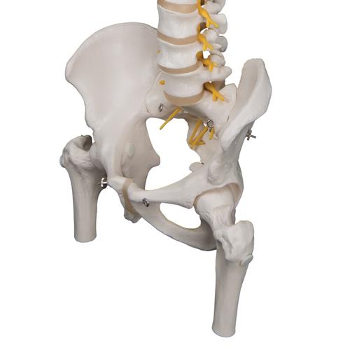
Anatomical Teaching Models Plastic Spinal Column Vertebrae Model Flexible Spine Model from www.3bscientific.com T10 is situated at the umbilicus. See lumbar spine anatomy diagram stock video clips. The first three vertebrae in the sacral have transverse processes which come together to form wide lateral wings called alae. This particular vertebra has a complete articular facet and the thoracic spinal nerves passes out under it. As the last of the lumbar vertebrae, the l5 vertebra bears more body weight than any of the other 23 vertebrae that sit atop it in the vertebral column. C5 injuries often maintain shoulder and biceps control, but have no control at the wrist or hand. The ribs connected to t11 and t12 at the bottom of the thoracic spine do not attach the sternum in front, but do provide protection for the kidneys in the back of the body. This level is also called the important transpyloric plane, since the pylorus of the stomach is at this level.
In addition, such a patient should recover hip extensors, knee extensors, and even ankle dorsiflexion.
Browse 20,526 human spine stock photos and images available, or search for human spine anatomy or human spine xray to find more great stock photos and pictures. Sacral nerves (s1 to s5) injuries generally result in some loss of functionin the hips and legs. These are bundled into specialized tracts that conduct impulses triggered by pressure, pain, heat, and other sensory stimuli or conduct motor impulses activating muscles and glands. Quadriplegia with normal arm function; The sacrum fits between the two hipbones connecting the spine to the pelvis located just below the lumbar vertebrae. Injuries at the thoracic level and below result in paraplegia, with the hands not affected. Lumbar nerves (l1 to l5) injuries generally result in some loss of function in the hips and legs. Extent of disability is determined by damage done to the t10 vertebra. The l4 and l5 disc, in between the l4 and l5 vertebrae, can herniate or degenerate, leading to possible leg pain (sciatica) and/or lower back pain. C5 injuries often maintain shoulder and biceps control, but have no control at the wrist or hand. See human spine stock video clips. The most distinctive characteristic of this bone is the strong odontoid process (dens) which rises perpendicularly from the upper surface of the body. See lumbar spine anatomy diagram stock video clips.
The t2 vertebra possesses facets that create joints with two of the ribs, thus helping to keep the thoracic spine far more stable than the cervical spine in the neck or the lumbar spine in the lower back. This diagram depicts picture of a human spine.human anatomy diagrams show internal organs, cells, systems, conditions, symptoms and sickness information and/or tips for healthy living. Five bones (abbreviated s1 through s5) fused into a triangular shape, form the sacrum. Often there is little or no voluntary control of bowel or bladder, but patients usually manage on their own with the use of special equipment. Some individuals have four lumbar vertebrae, while others have six.
Anatomical Teaching Models Plastic Spinal Column Vertebrae Model Deluxe Flexible Spine With Femur Heads from www.3bscientific.com The sacrum is located behind the pelvis. Often there is little or no voluntary control of bowel or bladder, but patients usually manage on their own with the use of special equipment. As part of the pelvic girdle, the sacrum forms the back wall of the pelvis and also forms joints at the hip bone called the sacroiliac joints. This diagram depicts picture of a human spine.human anatomy diagrams show internal organs, cells, systems, conditions, symptoms and sickness information and/or tips for healthy living. The sacrum is shaped different in males and females. In some subjects, the seventh cervical vertebra is associated with an abnormal pair of ribs, known as cervical ribs. Browse 20,526 human spine stock photos and images available, or search for human spine anatomy or human spine xray to find more great stock photos and pictures. The second vertebra in the thoracic spine is responsible for helping to support the rib cage.
Isolated against a white background.
The fifth lumbar vertebra is the most common site of spondylolysis and spondylolisthesis. The lumbar spine is highlighted by green colour. Quadriplegia with normal arm function; In some subjects, the seventh cervical vertebra is associated with an abnormal pair of ribs, known as cervical ribs. The third thoracic vertebrae is a small vertebra in the upper middle back that plays an integral role in supporting the rib cage. T4 syndrome is a relatively uncommon condition in which spinal injury at the t4 vertebra causes a set of symptoms including diffuse arm pain and pins and needles or numbness in the upper arm. Browse 20,526 human spine stock photos and images available, or search for human spine anatomy or human spine xray to find more great stock photos and pictures. The sacrum contains a series of four openings on each side through which the sacral nerves and blood vessels run. Comparatively speaking, t1 is the smallest of all thoracic vertebrae. If the fourth cervical vertebrae (c4) nerve root is also involved, pain is usually felt in the upper arms and shoulders, as well as the lower neck. Anterior cross section of spinal column revealing how vertebra fits round spinal cord. An injury in this area will most likely experience limited or complete loss of use of the muscles in the lower abdomen, buttocks, legs, and feet. Sacral nerves (s1 to s5) injuries generally result in some loss of function in the hips and legs.